Lithospermum erythrorhizon.or Arnebia euchroma. Onosma paniculatum. is also used as Zi cao in some parts of China.
  
紫草 Zǐ cǎo- "Purple herb" Redroot Gromwell, Sinkiang Arnebia  Family: Boraginaceae     
Arnebia is a rather soft plant and its root is called "soft purple herb"- Ruan zi cao. Lithospermum is harder and its root is accordingly referred to as "hard purple herb"- Ying zi cao.
PART USED: Dry root, whole plant.- harvested Spring or Autumn.
Nature: Cold    
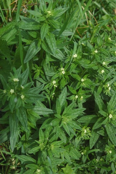
FLAVOR: Bitter, Sweet, Salty, Pungent, Pleasant, biting   CHANNEL: Heart, Liver
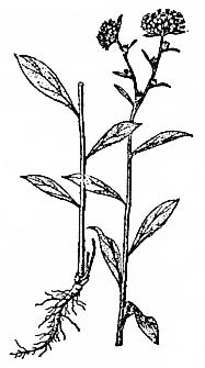
FUNCTIONS- Used as an emperor herb for treating children with measles and other Hot Toxic conditions
GROUP: Clearing Internal Heat- Cool Blood- Detoxifies
1. Cool Heat and Detoxifies. Clears fevers and Cools Blood.[1]
2. Detoxifies and lubricates the intestines.[1] Moistens the Intestines and unblocks the bowels.[4]
3. Clears Damp Heat from the skin.[4]
ACTIONS
INDICATIONS
1. Relieves Fire toxicity while venting rashes.[4] Measles preventive.[1] Encourages measles or chicken pox to surface.[4] Neutralize toxins- bring out measles.[3] Often used for fire toxin with very dark purple rashes.[4]
2. Burns, knife cuts and bleeding.[1]
3. Oozing dermatitis, abscesses.[1]
4. Constipation-[1,3] Hot symptoms- particularly for Yang children.[3] Heat in the Blood accompanied by constipation.[4]
5. Preventative herb for season.[3]
Topically for Damp Heat skin lesions or vaginal itching.[4] Also used for burns.[4]
CONTRAINDICATIONS: Measles that are erupting normally.[4] Use with caution in cases of Spleen or Stomach deficiency as it has a mild laxative effect.[4]
COMBINATIONS
PREPARATIONS: Decoction- whole plant 9-15 g.[1] Dry Root.[2] 3-9 g.[3,4] Often made into an ointment for external use.[4]

HABITAT: Found growing on sunny hillsides.
DESCRIPTION: Perennial herb. Stem: erect, height reaching 60 cm, pubescent with white hairs. Leaves: alternate, broadly lanceolate, apexes acute, bases cuneate, margins intact. Flowers: in autumn, white terminal flowers appear to form racemose inflorescences. Nut: small, ovoid, light brown
References
Inner Path can not take any responsibility for any adverse effects from the use of plants. Always seek advice from a professional before using a plant medicinally.

Constituents

Research

Shikonin, an ingredient of Lithospermum erythrorhizon, inhibits angiogenesis in vivo and in vitro.
Hisa T, Kimura Y, Takada K, Suzuki F, Takigawa M.
Abstract
Angiogenesis is critical for tumor growth and inflammation. Shiunko is a Chinese herbal ointment used for the treatment of burns in Japan. Its main ingredient is the root of Lithospermum erythrorhizon, which had been used for treating tumors and inflammation in China since the 5th century. We report here that shikonin, the main chemical ingredient of L. erythrorhizon is a novel inhibitor of angiogenesis. It inhibited tumor necrosis factor-alpha-induced and B16 melanoma-induced angiogenesis in mice and normal developmental angiogenesis in the yolk-sac membranes of chick embryos. Shikonin also inhibited proliferation and migration of endothelial cells in culture and network formation by endothelial cells on Matrigel in vitro. The dose-responsive study suggests that the mechanism of this inhibitory effect on angiogenesis involves the prevention of network formation by endothelial cells via blocking integrin alpha v beta 3 expression.
PMID: 9615720 Anticancer Res. 1998 Mar-Apr;18(2A):783-90. ncbi.nlm.nih.gov

Gromwell (Lithospermum erythrorhizon) Supplementation Enhances Epidermal Levels of Ceramides, Glucosylceramides, β-Glucocerebrosidase, and Acidic Sphingomyelinase in NC/Nga Mice
Jungmin Kim and Yunhi Cho
Abstract
We have previously reported that dietary gromwell (Lithospermum erythrorhizon; LE) prevents the development of atopic dermatitis (AD) with increased epidermal levels of total ceramide (Cer), the major lipid maintaining epidermal barrier. In this study, we investigated whether the increased level of total Cer induced by dietary LE would be related to the altered metabolism of glucosylceramide (GlcCer) and sphingomyelin (SM), two major precursor lipids in Cer generation. NC/Nga mice, an animal model of AD, were fed a control diet (group CA: atopic control) or a diet with 70% ethanol LE extracts (1% in diet; group LE) for 10 weeks. Individual species of Cer, GlcCer, and SM were analyzed by high-performance thin layer chromatography. In the epidermis of group CA, total Cer (including Cer2 and Cer5–7) and total GlcCer (including GlcCer-B/C/D) were significantly reduced; these levels in group LE were increased to levels similar to the normal control group of BALB/c mice (group C). In addition, protein expressions and activities of β-glucocerebrosidase (β-GlcCer'ase) and acidic sphingomyelinase (aSMase), enzymes for GlcCer or SM hydrolysis, respectively, were increased in group LE. However, alterations of Cer1, Cer3/4, GlcCer-A, and all SM species (including SM1–3) were not significant among groups C, CA, and LE. Dietary gromwell increases GlcCer-B/C/D, and further enhances the generation of Cer2 and Cer5–7 with high protein expressions and activities of β-GlcCer'ase and aSMase.
J Med Food. 2013 Oct; 16(10): 927–933. doi: 10.1089/jmf.2012.2734 PMCID: PMC3806403 ncbi.nlm.nih.gov

Lithospermum erythrorhizon extract protects keratinocytes and fibroblasts against oxidative stress.
Yoo HG, Lee BH, Kim W, Lee JS, Kim GH, Chun OK, Koo SI, Kim DO.
Abstract
Oxidative stress damages dermal and epidermal cells and degrades extracellular matrix proteins, such as collagen, ultimately leading to skin aging. The present study evaluated the potential protective effect of the aqueous methanolic extract obtained from Lithospermum erythrorhizon (LE) against oxidative stress, induced by H2O2 and ultraviolet (UV) irradiation, on human keratinocyte (HaCaT) and human dermal fibroblast-neonatal (HDF-n) cells. Exposure of cells to H2O2 or UVB irradiation markedly increased oxidative stress and reduced cell viability. However, pretreatment of cells with the LE extract not only increased cell viability (up to 84.5%), but also significantly decreased oxidative stress. Further, the LE extract downregulated the expression of matrix metalloproteinase-1, an endopeptidase that degrades extracellular matrix collagen. In contrast, treatment with the LE extract did not affect the expression of procollagen type 1 in HDF-n cells exposed to UVA irradiation. Thirteen phenolic compounds, including derivatives of shikonin and caffeic acid, were identified by ultrahigh-performance liquid chromatography-electrospray ionization-tandem mass spectrometry. These results suggest that LE-derived extracts may protect oxidative-stress-induced skin aging by inhibiting degradation of skin collagen, and that this protection may derive at least in part from the antioxidant phenolics present in these extracts. Further studies are warranted to determine the potential utility of LE-derived extracts in both therapeutic and cosmetic applications.
PMID: 25136892 DOI: 10.1089/jmf.2013.3088 J Med Food. 2014 Nov;17(11):1189-96. doi: 10.1089/jmf.2013.3088. Epub 2014 Aug 19. ncbi.nlm.nih.gov

In vitro and in vivo anticancer effects of Lithospermum erythrorhizon extract on B16F10 murine melanoma.
Rajasekar S, Park DJ, Park C, Park S, Park YH, Kim ST, Choi YH, Choi YW.
Abstract
ETHNOPHARMACOLOGICAL RELEVANCE:
Lithospermum erythrorhizon has long been used in traditional Asian medicine for the treatment of diseases including skin cancer. In this study, hexane extract from the roots of Lithospermum erythrorhizon (LEH) was chemically characterized and its anticancer activity was tested against the most aggressive form of skin cancer.
MATERIALS AND METHODS:
The in vitro anticancer studies viz. cell growth, cell cycle and apoptosis, and the expression of tumor regulating proteins were analyzed against B16F10 melanoma cells. In addition, C57BL/6 mice models were used to evaluate the in vivo anticancer potential of LEH. Mice were intraperitoneally injected with LEH at doses of 0.1 and 10mg/kg every 3 days. The tumor inhibition ratio was determined after 21 days of treatment and the histopathological analyses of the tumor tissues were compared. Further, LEH was purified and its active compounds were structurally elucidated and identified by NMR spectra and quantified by HPLC analyses.
RESULTS:
LEH effectively inhibits the growth of melanoma cells with an IC(50) of 2.73μg/ml. Cell cycle analysis revealed that LEH increased the percentage of cells in sub-G1 phase by dose dependent manner. LEH exhibited down regulation of anti-apoptotic Bcl-2 family proteins and up regulation of apoptotic Bax protein expression. Importantly, LEH induced cleavage of poly (ADP-ribose) polymerase (PARP) and activated the caspase cascade (caspase 3) with this cleavage mediating the apoptosis of B16F10 cells. LEH treatment at a dose of 10mg/kg for 21 days in experimental mice implanted with tumors resulted in significant reduction of the tumor growth (43%) and weight (36%). Histopathology analysis of LEH treated tumor tissues showed evidence of increased necrotic cells in a concentration dependent manner. Meanwhile, five naphthoquinone compounds [Shikonin (1); Deoxyshikonin (2); β-Hydroxyisovalerylshikonin (3); Acetylshikonin (4) and Isobutyrylshikonin (5)] were purified from LEH and responsible for its anticancer activity.
CONCLUSION:
LEH induced apoptosis in B16F10 cells by activation of caspase 3 and inducing sub-G1 cell cycle arrest. LEH exhibited both in vitro and in vivo anticancer activity. Shikonin derivatives in the LEH are responsible for the anticancer activity.
PMID: 22995444 DOI: 10.1016/j.jep.2012.09.017  J Ethnopharmacol. 2012 Nov 21;144(2):335-45. doi: 10.1016/j.jep.2012.09.017. Epub 2012 Sep 17. ncbi.nlm.nih.gov