Cervus nippon鹿茸
Lù róng Sika
deer, Spotted deer
Family: Cerridae PART USED:Velvet from
precalcified antler. Velvet is the outermost layer of skin covering
a growing antler, and is hair like in appearance and texture.
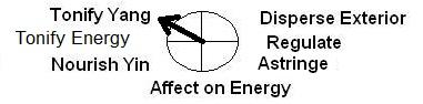
FLAVOR: Sweet, salty CHANNEL: Kidney, Liver FUNCTIONS GROUP: Replenishing Yang-
Tonic
1. Tonify Yang.
2. Nourish semen and Blood.[1,2] Benefit
semen.
3. Strengthen the tendons and bones.[1,2]
Tone up marrow, strengthen bones. INDICATIONS
1. Kidney Yang deficicency manifested as cold limbs, fatigue, backache.
Impotence. Seminal emission and frequent micturition, enuresis, sterility, metrorrhagia
or leukorrhagia. Premature ejaculation. Fear of cold with weakness.
2. Kidney deficiency with shortness of breath and dyspnea.
3. Yang deficiency and insufficiency of essence and blood manifested as flaccidity
of extremities, or maldevelopment, delayed walking, delayed teeth eruption and
delayed closure of fontanelle in infants.
4. Blood deficiency, dizziness, vaginal bleeding due to Cold and deficient conditions.
Unhealed skin lesions. COMPARISON: Lu rong with Lu jiao
Lu rong is the velvet havested from the antler
Lu jiao is the discarded horns- which are grown each year
Lu Rong and Lu Jiao are very different, from their chemical components to their
pharmacological functions. Lu Rong contains 50% amino acids, while Lu Jiao contains
50-60% calcium phosphate.[3] PREPARATIONS:Pill
or powder 0.25-0.5 bid or tid.[2]
Pilose antler of a young stag 1-2.5 g.[1]
[1] A Complete English Dictionary of Medicinal
Terms in Chinese Acupuncture and Herbalism 1981- Henry Lu Chinese Foundations
of Natural Health- The Academy of Oriental Heritage, Vancouver, Canada.
[2] Medicated Diet of Traditional Chinese Medicine- Chief Editor- Hou Jinglun.
Associate Editors- Zhao Xin, Li Weidong, Liu Jianxin, Geng Chun-e, Li Guohua,
Li Shaohua. Geijing. Science & Technology Press 1994. ISBN 7-5304-1735-5/R.
309.(Lu).
[3] safflower.com.au Images
1. en.wikipedia.org
by Quartl CC BY-SA
3.0
2. englishtaobao.net
Inner Path can not take any responsibility for any adverse effects from the
use of plants. Always seek advice from a professional before using a plant medicinally. Research
First Evidence that Sika Deer (Cervus nippon) Velvet Antler Extract
Suppresses Migration of Human Prostate Cancer Cells
YuJiao Tang, Byong-Tae Jeon, Yanmei Wang, Eun-Ju Choi, Yon-Suk Kim, Jin-Woo
Hwang, Pyo-Jam Park, Sang Ho Moon, and Eun-Kyung Kim Abstract
Deer velvet antler (DVA) is one of the most popular medicines in China. Numerous
studies have demonstrated that velvet antler possess biological effects. However,
data regarding its anti-migration activity on prostate cancer is scarce. In
this study, we investigated the inhibitory effect of top DVA (T-DVA) on the
expression of prostate-specific antigen (PSA) and migration-related genes in
the human prostate cancer cell, LNCaP. The T-DVA down-regulated the expression
of PSA. In addition, the RadiusTM assay revealed that T-DVA inhibited the migration
behavior of prostate cancer cells. Furthermore, the expression of matrix metalloproteinase
(MMP)-9 and vascular endothelial growth factor (VEGF) was also decreased with
T-DVA. On the contrary, T-DVA increased the tissue inhibition of metalloproteinase
(TIMP)-1 and (TIMP)-2. Taken together, our findings indicate that the T-DVA
possesses anti-migration activity on prostate cancer cells. This is the first
study of DVA to report the anti-migration activity on prostate cancer.
Korean J Food Sci Anim Resour. 2015; 35(4): 507–514.
Published online 2015 Aug 31. doi: 10.5851/kosfa.2015.35.4.507 PMCID: PMC4662134
ncbi.nlm.nih.gov
Prevention and therapeutic effects of sika deer velvet collagen hydrolysate
on osteoporosis in rats by retinoic acid. [Article in Chinese]
Li Y, Zhao Y, Sun X, Qu X. Abstract
The objective was to evaluate the preventive and therapeutic effects of the
collagen hydrolysate extracted from Sika deer velvet (CSDV) on osteoporosis
rats induced by retinoicacid. Histomorphometric indices and serum biochemical
parameters were measured in osteoporosis rats treated with/without antler collagen
and in sham-operated rats. Our results were as follows: compared with the osteoporosis
group, significant elevation in the levels of bone mineral density (BMD), Ca,
P and static histomorphometric indexes and biomechanical properties, but reduction
in the level of serum alkaline phosphatase (ALP) were observed in antler collagen-treated
groups. However, the above function with the collagenase solution velvet material
varied with the different doses. In conclusion, the extracted collagen is found
to play a role in the prevention and treatment of osteoporosis rats by retinoic
acid.
PMID: 20545204 Zhongguo Zhong Yao Za Zhi. 2010 Mar;35(6):759-62. ncbi.nlm.nih.gov
Effects of deer velvet extract from Formosan sika deer on the embryonic
development and anti-oxidative enzymes mRNA expression in mouse embryos.
Cheng SL, Lai YL, Lee MC, Shen PC, Liu SS, Liu BT. Abstract
ETHNOPHARMACROLOGICAL RELEVANCE:
The deer velvet or its extracts has been widely used in clinic. It has been
used in promoting reproductive performances and treating of oxidation and aging
process. The aim of this study is to investigate the effects of velvet extract
from Formosan sika deer (Formosan sika deer; Cervus nippon taiouanus, FSD) velvet
on mouse embryonic development and anti-oxidant ability in vitro.
MATERIALS AND METHODS:
Mouse 4-cells embryos were divided into 16 groups for 72 h in vitro incubation.
The embryonic development stages and morphology were evaluated every 12h in
experimental period. The quantitative real time PCR was used to measure the
CuZn-SOD, GPx and CAT mRNA expression of the blastocysts.
RESULTS:
The 4-cells embryos of hydrogen peroxide (HP) groups did not continue developing
after oxidant stress challenged. The blastocyst developmental rate (90.0-90.4%,
P>0.05) and normal morphological rate (84.4-85.1%, P>0.05) of the 1% and
2% DV extract groups were similar to those in the control group (90.7% and 88.8%,
respectively). The embryos challenged by HP (5, 10 and 25 μM) and subsequently
incubated in mHTF medium with 1% and 2% of deer velvet (DV) extracts were able
to continue development; the blastocyst developmental rate of these groups were
similar to that in the control group. The relative mRNA expression of the focused
anti-oxidative enzymes in the mouse embryos did not significantly differ among
the designed DV treatment groups (P>0.05).
CONCLUSION:
The FSD velvet extract in adequate concentration could promote anti-oxidative
enzymes mRNA expression followed the challenge of hydrogen peroxide, relieve
the mouse embryo under oxidative stress, and maintain the blastocyst developmental
ability in vitro.
PMID: 24732110 DOI: 10.1016/j.jep.2014.04.006 J Ethnopharmacol. 2014 Jul
3;154(3):600-5. doi: 10.1016/j.jep.2014.04.006. Epub 2014 Apr 13. ncbi.nlm.nih.gov
Sika Deer Antler Collagen Type I-Accelerated Osteogenesis in Bone Marrow
Mesenchymal Stem Cells via the Smad Pathway
Na Li, Min Zhang, Gregor P. C. Drummen, Yu Zhao, Yin Fen Tan, Su Luo, and Xiao
Bo Qu Abstract
Deer antler preparations have been used to strengthen bones for centuries. It
is particularly rich in collagen type I. This study aimed to unravel part of
the purported bioremedial effect of Sika deer antler collagen type I (SDA-Col
I) on bone marrow mesenchymal stem cells. The results suggest that SDA-Col I
might be used to promote and regulate osteoblast proliferation and differentiation.
SDA-Col I might potentially provide the basis for novel therapeutic strategies
in the treatment of bone injury and/or in scaffolds for bone replacement strategies.
Finally, isolation of SDA-Col I from deer antler represents a renewable, green,
and uncomplicated way to obtain a biomedically valuable therapeutic.
Evidence-Based Complementary and Alternative Medicine
Volume 2016 (2016), Article ID 2109204, 13 pages http://dx.doi.org/10.1155/2016/2109204
Received 30 September 2015; Accepted 6 December 2015 hindawi.com
 in the Lüneburg Heath wildlife park, Germany.")
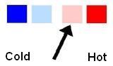
 FLAVOR: Sweet, salty CHANNEL: Kidney, Liver